BONE SUBSTITUTES
ANIMUS HUMAN BONE GRAFT
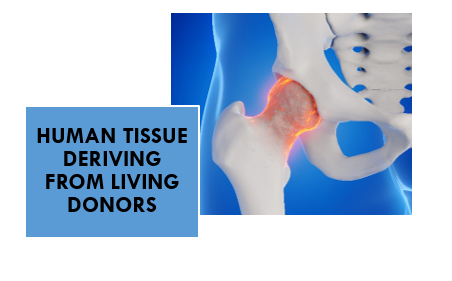
Animus is an allograft , derived from human living donors,is a key material in dental regenerative procedures. Offering osteoconductive and, in some forms, osteoinductive properties, ANIMUS serve as a versatile option for bone augmentation in implantology and periodont
Bone loss in the oral cavity due to trauma, infection, periodontal disease, or tooth extraction often necessitates regenerative procedures to restore lost volume. When autogenous bone is not viable or desirable, allograft bone—graft material obtained from a human donor—provides an effective alternative. ANIMUS avoid donor site morbidity, are widely available, and come in various forms tailored for specific dental applications.
Source
Allograft bone is obtained from LIVING human donors, screened and processed through certified tissue banking procedure in accordance with EATB(European Association of Tissue Banks) and AATB (American Association of Tissue Banks) standards.
Allograft bone is obtained from LIVING human donors, screened and processed through certified tissue banking procedure in accordance with EATB(European Association of Tissue Banks) and AATB (American Association of Tissue Banks) standards.
Processing Techniques
To ensure safety and preserve graft function, processing includes:
- Demineralization (for exposing growth factors like BMPs in DMBM)
- Freeze-drying (lyophilization) to preserve structure and shelf life
- Chemical sterilization for microbial safety
- Decellularization to reduce immunogenicity
- Labeling and Packaging
- Sterilization gamma radiation
Donor bone is rigorously tested for HIV, hepatitis, and other transmissible diseases before and after processing
Biological Properties
Osteoconduction
ANIMUS act as a biocompatible scaffold, allowing for the migration of osteogenic cells and new bone formation.
Osteoinduction
Demineralized bone matrix (DBM) retains non-collagenous proteins such as bone morphogenetic proteins (BMPs), which may stimulate the recruitment and differentiation of osteoprogenitor cells.
Biocompatibility
ANIMUS integrates with host tissue and exhibit low immunogenicity, though rare inflammatory reactions have been reported.
Clinical Applications
Socket Preservation
ANIMUS is placed post-extraction to maintain ridge dimensions and minimize resorption.
Ridge Augmentation
Used in horizontal or vertical augmentation procedures to recreate bone volume for implant placement.
Sinus Floor Elevation
Effectively used in both lateral and transcrestal sinus lifts, often in combination with xenografts for volume maintenance.
Periodontal Regeneration
ANIMUS is used in intrabony and furcation defects for periodontal regeneration.
Peri-Implant Defect Management
Fill defects around implants to enhance osseointegration and support long-term function.
- Eliminates need for donor site (no second surgical site)
- Wide availability and shelf-stable
- Versatile (available in multiple forms and particle sizes)
- Biologically active (especially DBM)
- Proven clinical efficacy
Clinical Evidence
Numerous studies support the use of ANIMUS bone in dentistry:
- A 2016 systematic review found comparable outcomes in implant survival between sites grafted with allograft and autograft bone [1].
- DBM studies show enhanced periodontal regeneration when combined with membranes or growth factors [2].
- Long-term clinical success reported in ridge preservation and sinus lift cases using mineralized freeze-dried bone allografts (FDBA) [3].
References
- Wallace SC, Froum SJ. (2003). Effect of maxillary sinus augmentation on the survival of endosseous dental implants. A systematic review. Ann Periodontol. 8(1):328–343.
- Mellonig JT. (2000). Human histologic evaluation of a xenograft in the treatment of periodontal osseous defects. Int J Periodontics Restorative Dent. 20(6):543–549.
- Misch CE, Dietsh-Misch F. (1993). The use of allograft bone in preprosthetic surgery. Oral Health. 83(5):15–23.
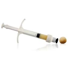
ANIMUS Demineralized Bone Matrix (DBM) Putty
Demineralized bone matrix (DBM) putty is a biologically active, moldable bone graft material widely used in orthopedic, neurosurgery, oral and maxillofacial regenerative procedures. Derived from human bone, DBM contains collagen and growth factors that promote bone healing.
The regeneration of bone in the oral cavity is fundamental to successful dental implant placement, periodontal therapy, and ridge preservation. When autologous bone grafting is not feasible, ANIMUS allograft material and ANIMUS Demineralized Bone Matrix (DBM) serve as effective alternatives. ANIMUS Demineralized Bone Matrix in putty form offers both osteoconductive scaffolding and potential osteoinductive activity, with superior handling properties that make it suitable for diverse clinical settings.
ANIMUS Demineralized Bone Matrix (DBM)
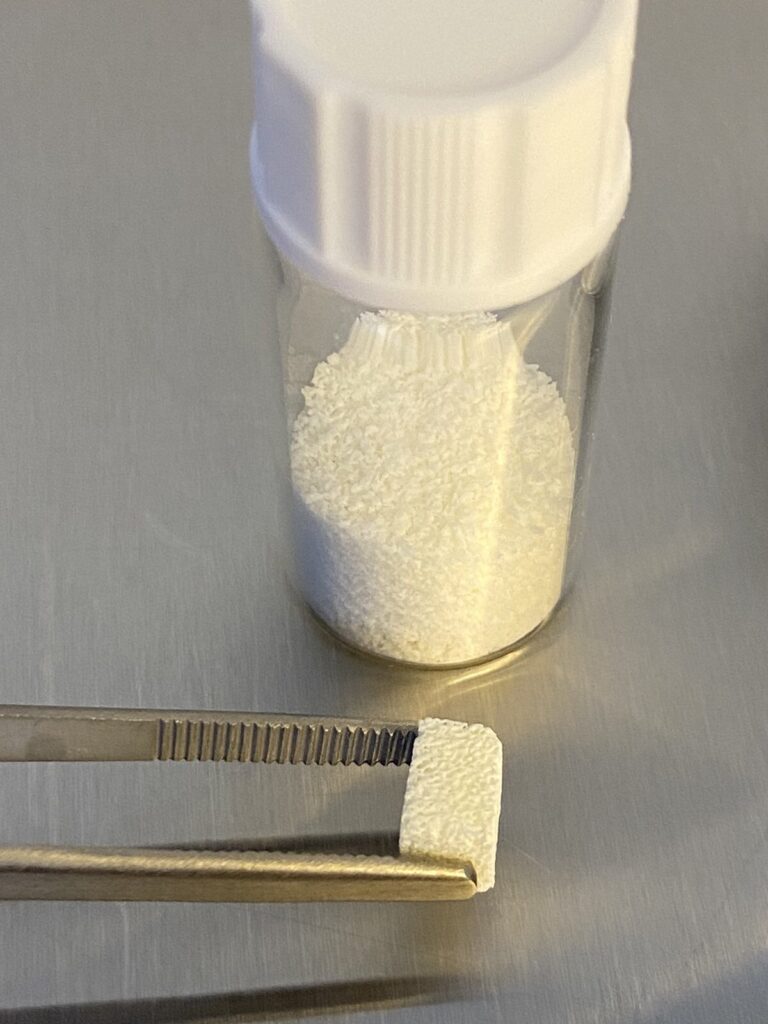
ANIMUS Demineralized Bone Matrix is an allograft derived from the cortical and cancellous bone of screened human donors. The bone is decalcified using acid extraction to remove the inorganic mineral phase (primarily hydroxyapatite), while preserving:
- Type I collagen
- Non-collagenous proteins
- Growth factors, such as:
- Bone Morphogenetic Proteins (BMPs)
- Transforming Growth Factor-beta (TGF-β)
These components are key to DBM’s osteoinductive properties, making it unique among allograft materials.
ANIMUS Demineralized Bone Matrix DBM putty combines this matrix with a biocompatible carrier to improve handling and ensure consistency during placement.
Mechanism of Action
DBM putty supports bone regeneration through:
Osteoconduction
ANIMUS Demineralized Bone Matrix Acts as a scaffold that allows for cellular ingrowth and vascularization.
Osteoinduction
ANIMUS Demineralized Bone Matrix Stimulates mesenchymal stem cells to differentiate into osteoblasts via growth factors like BMPs—though potency varies by product and donor.
Remodeling
Once implanted, the material is gradually replaced by native bone through a natural remodeling process.
Clinical Applications in Dentistry
Ridge Preservation
Placed in extraction sockets to minimize bone resorption and support future implant placement.
Periodontal Regeneration
Used to fill intrabony defects and furcations in combination with barrier membranes.
Sinus Augmentation
Serves as a space-filling material in both lateral and transcrestal sinus lift procedures, often combined with xenografts for volume stability.
Peri-implant Defects
Applied to fill gaps or dehiscences around immediate or delayed implants.
Cystic Cavity Fill
Used to fill osseous defects following cyst enucleation or apicoectomy.
Advantages
- Osteoinductive potential due to growth factor content
- Excellent handling: moldable, adaptable to defect shape
- No donor site morbidity
- Shelf-stable and available in various volumes and concentrations
- Combines well with other grafts (e.g., bovine bone, autograft, PRF)
Clinical Performance and Evidence
Several studies support the effectiveness of DBM putty:
- Histologic evidence confirms new bone formation and vascular ingrowth at grafted sites within 3–6 months [1].
- Periodontal studies report improved clinical attachment and defect fill compared to open flap debridement alone [2].
- In sinus lift procedures, DBM putty combined with xenografts has demonstrated predictable bone regeneration and implant integration [3].
ANIMUS Demineralized Bone Matrix Putty is a versatile and biologically active grafting material that supports bone regeneration in a wide range of dental procedures. With its ease of handling and osteoinductive potential, DBM putty is especially effective in non-load-bearing applications such as socket preservation and periodontal regeneration. While variability in performance exists, careful product selection and case planning can yield predictable and successful outcomes.
References
- Schwartz Z, et al. (1996). Histological evaluation of demineralized freeze-dried bone allograft in socket preservation. J Periodontol, 67(9), 918–926.
- Mellonig JT. (1995). Clinical evaluation of demineralized bone matrix in periodontal defects. Int J Periodontics Restorative Dent, 15(5), 469–481.
- Wallace SC, et al. (2005). The use of DBM in sinus elevation: A retrospective study. Implant Dent, 14(2), 128–135.
ZOE Bovine Bone Graft
ZOE Bovine Bone Graft is one of the most widely used xenografts in dental and maxillofacial surgery. It’s osteoconductive properties, safety profile, and structural similarity to human bone it them ideal for a variety of regenerative procedures, including socket preservation, ridge augmentation, and sinus lifts.
Bone regeneration is critical in restorative and implant dentistry. Adequate bone volume is essential for successful implant placement and long-term function. When natural bone healing is insufficient, graft materials are required to support regeneration ZOE Bovine Bone Graft is a xenograft derived from the femoral bone of cattle, presents very high biological and mechanical compatibility with human bone.
Origin
Bovine bone is harvested from healthy cattle, primarily from cortical or cancellous bone. The structure and mineral composition are closely related to that of human bone.
Processing Methods
To eliminate any risk of disease transmission and immunogenicity, bovine bone undergoes rigorous processing, including:
- Deproteinization to remove organic components and antigens
- Demineralization (optional, for certain products) to expose bioactive proteins
- Labeling and Packaging
- Sterilization by high heat, gamma irradiation, and chemical agents
This results in an anorganic, sterile, porous matrix which serves as a scaffold for new bone growth.
High Biological Properties
Osteoconduction
ZOE Bovine Bone Graft act as a biocompatible scaffold, supporting the migration, adhesion, and proliferation of osteoblasts and integrate well into host bone over time.
Slow Resorption
Due to it’s high crystallinity and low solubility, ZOE Bovine Bone Graft
resorb slowly, maintaining volume over extended healing periods.
Porosity and Structure
The porous architecture mimics natural cancellous bone, facilitating vascular infiltration and bone regeneration.
Clinical Applications
Socket Preservation
Placed in extraction sites to prevent alveolar ridge resorption, maintaining ideal volume for future implant placement.
Ridge Augmentation
Used to restore horizontal and vertical bone deficiencies prior to implant placement.
Sinus Lift Procedures
Serves as a scaffold in lateral and crestal sinus augmentation, allowing for predictable bone formation beneath the Schneiderian membrane.
Peri-Implant Defects
Fills dehiscence and fenestration defects around implants to promote osseointegration.
Periodontal Regeneration
Combined with membranes for treating infrabony periodontal defects in select cases.
Advantages
- High biocompatibility with minimal immunogenic response
- Volume stability due to slow resorption
- Proven clinical track record across thousands of cases
- Readily available and cost-effective compared to autografts
Clinical Outcomes and Evidence
- High biocompatibility with minimal immunogenic response
- Volume stability due to slow resorption
- Proven clinical track record across thousands of cases
- Readily available and cost-effective compared to autografts
References
- Artzi Z, et al. (2005). Histological comparison of anorganic bovine bone and autogenous bone in sinus augmentation. Clin Oral Implants Res, 16(4), 435–444.
- Traini T, et al. (2007). Long-term results of implants placed in sinuses grafted with anorganic bovine bone. J Periodontol, 78(5), 955–961.
- Jensen SS, Terheyden H. (2009). Bone augmentation procedures in localized defects in the alveolar ridge: clinical results with different bone grafts and bone-substitute materials. Int J Oral Maxillofac Implants, 24 Suppl:218–236.
ZOE +HYAL 4% Bovine Bone Graft
Hyaluronic acid (HA), a naturally occurring glycosaminoglycan, has emerged as a valuable additive in bone grafting procedures, including dental and maxillofacial surgeries. When incorporated into graft or used as a carrier, HA offers biological and handling benefits that support bone healing and regeneration.
Here’s a breakdown of the advantages of ZOE +HYAL 4% Bovine Bone Graft:
✅ 1. Enhanced Wound Healing
- HA promotes early soft tissue healing by stimulating fibroblast migration and angiogenesis (new blood vessel formation).
- It acts as a scaffold for epithelial and connective tissue cells, improving the healing of both soft and hard tissues post-surgery.
✅ 2. Osteoinductive Support
- While not inherently osteoinductive, ZOE +HYAL 4% Bovine Bone Graft enhances the biological activity attracting and retaining growth factors such as:
- Bone morphogenetic proteins (BMPs)
- Platelet-derived growth factor (PDGF)
- This may accelerate osteoblast recruitment and differentiation, aiding bone formation.
✅ 3. Improved Stability and Handling
- HA adds viscosity and cohesiveness to granules , making them easier to mold and apply.
- Acts as a binder, reducing graft particle migration or washout from the defect site.
- ZOE +HYAL 4% Bovine Bone Graft can help maintain graft shape and volume during healing.
✅ 4. Anti-inflammatory and Antibacterial Properties
- ZOE +HYAL 4% Bovine Bone Graft helps modulate inflammation, minimizing post-operative swelling and discomfort.
- It may have mild antimicrobial effects, lowering infection risk at the graft site.
✅ 5. Promotion of Angiogenesis
- ZOE +HYAL 4% Bovine Bone Graft encourages the formation of capillaries in and around the grafted area, improving vascularization, which is critical for graft integration and new bone formation.
✅ 6. Biodegradability and Biocompatibility
- ZOE +HYAL 4% Bovine Bone Graft is naturally resorbed by the body and does not interfere with bone remodeling.
- Its non-immunogenic nature ensures excellent biocompatibility, even in immunocompromised or medically complex patients.
Clinical Applications in Dentistry
- Socket preservation
- Ridge augmentation
- Periodontal defect regeneration
- Sinus augmentation
- Peri-implant defect repair
🧠 Summary Table: ZOE +HYAL 4% Bovine Bone Graft
Advantage | Clinical Benefit |
Hydrophilic & viscous | Improved handling of particulate grafts |
Promotes angiogenesis | Faster vascularization and integration |
Supports cell migration | Aids in osteoblast and fibroblast activity |
Anti-inflammatory | Reduces post-op swelling and discomfort |
Biocompatible and biodegradable | No long-term foreign body response |
Growth factor retention | Enhances osteogenic potential of graft site |